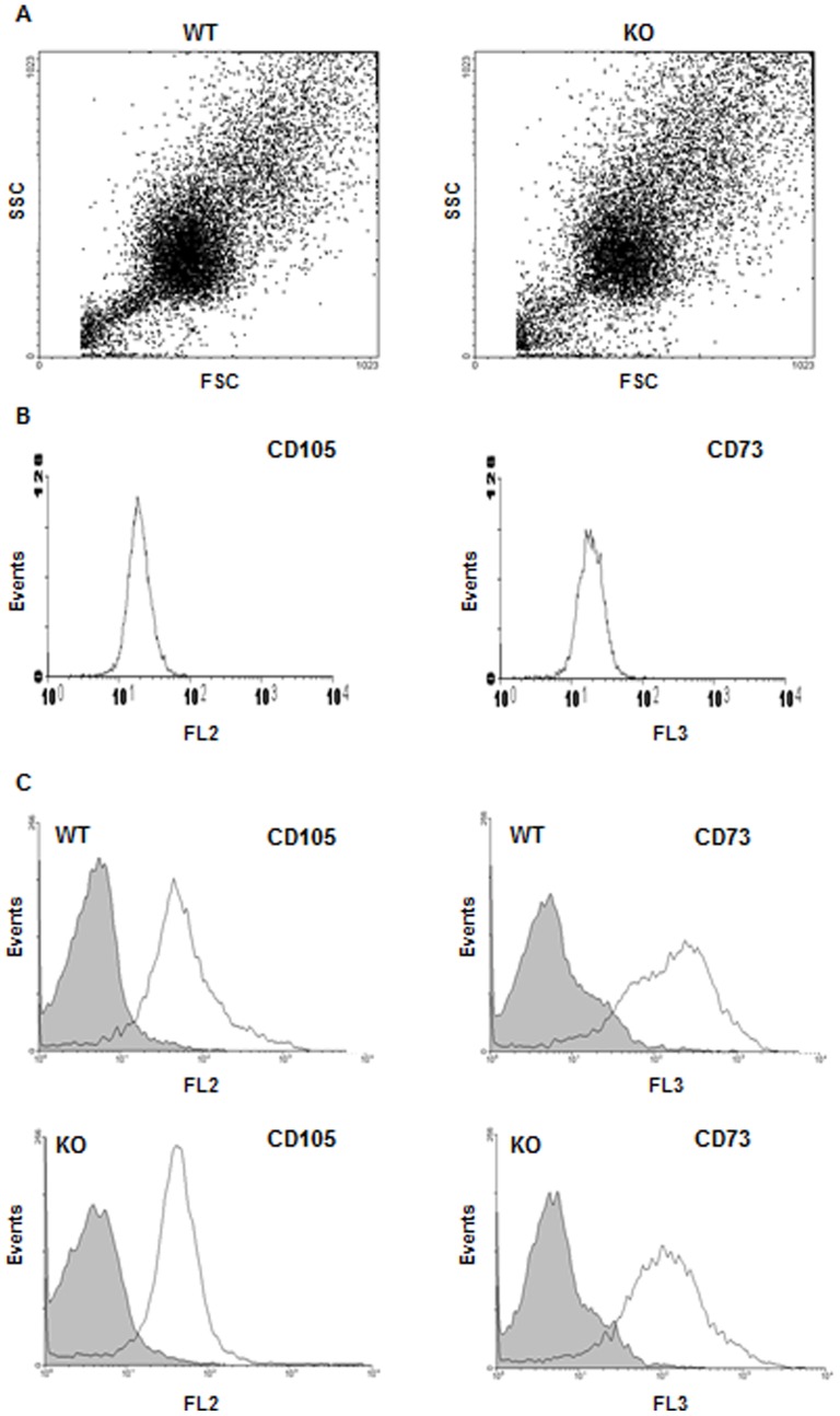
Figure 6. Phenotypic profile of bone marrow-derived MSC from WT and CD36KO mice.
Flow cytometry analysis of MSC isolated from bone marrow of WT and CD36KO mice was performed on cells freshly isolated and adherent cells after 11 days of culture using CD105-PE and CD73-PerCP antibodies. A) Forward scatter (FSC) and side scatter (SSC) of the MSC population from bone marrow of WT and CD36KO mice at day 11 of culture. B) CD105-PE and CD73-FITC staining of the mesenchymal cell line C3H10T1/2. C) CD105-PE and CD73-FITC fluorescence for freshly isolated bone marrow cells (grey) and adherent cells after 11 days of culture (clear) from WT and CD36KO mice.

