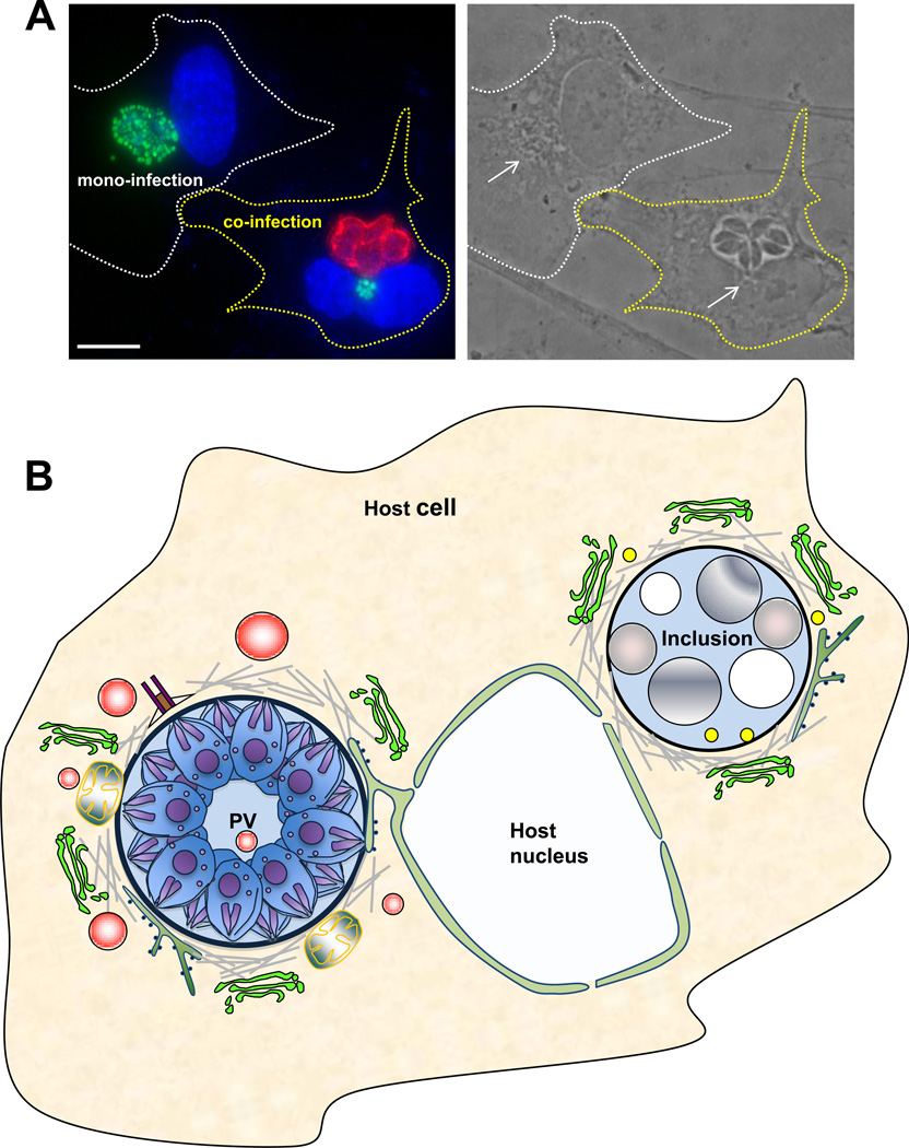
Figure 3. Co-infection of mammalian cells with Toxoplasma gondii and Chlamydia trachomatis, and host cell interactions with the PV and the inclusion during co-infection.
A. A phase and immunofluorescence image of a mono- and a co-infected epithelial cellsshowing the poor development of the inclusion (stained for EF-Tu in green; arrow) during a 24-h co-infection while the parasites (stained for GRA7 in red) develop normally. Both the inclusion and the PV are located in the perinuclear region of the host cell. Scale bar is 10 µm. B. A schematic representation of a PV and an inclusion occupying the same cell for 24-h summarizing their interaction with host cell structures. Similarly as in a mono-infection, the PV is anchored to the host nuclear envelope, associates with the host MTOC, microtubules, ER, mitochondria and attracts host Golgi fragments and endocytic organelles (in red) that are further delivered into the PV. In contrast, inclusions growing in a PV-containing cell shift to a stress-induced persistence state with large aberrant bodies despite its association with host microtubules (in the absence of the MTOC), Golgi fragments, ER and CERT vesicles, and the presence of lipid bodies in the vacuole.

