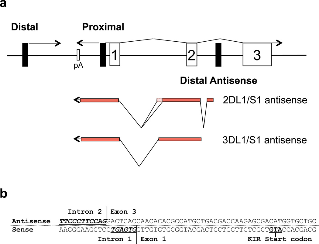
Figure 1.
Identification of KIR distal antisense transcripts. (a) A schematic of the organization of the 5’ region of the KIR genes is shown. Black rectangles represent promoter elements, and numbered rectangles represent exons. Lines represent KIR transcripts with their orientation indicated by arrows. The exon structure of the KIR2DL1/S1 and KIR3DL1/S1 distal antisense transcripts is indicated below. The additional exon sequence found in the KIR2DL1 alternative transcript (GenBank: GQ422373) is indicated by the dotted rectangle. (b) The nucleotide sequence of the region containing the KIR2DL1 distal antisense intron 2/exon 3 splice junction and the exon 1/intron 1 splice junction of the KIR2DL1 coding transcript is shown. The antisense strand is shown on top with the complementary coding sense strand shown on the bottom. Consensus splice acceptor (antisense) and splice donor (sense) sequences are underlined in bold.

