Figure 4.

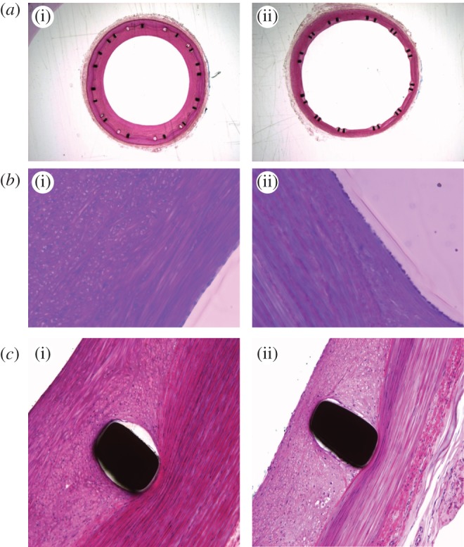
(a) Low magnification fuchsin stained cross sections of porcine common carotid arteries with (i) straight-centreline stent implanted and (ii) helical-centreline stent implanted. (b) Same cross sections as in figure 4a (40× magnification). An endothelial cell layer is visible in both (i) and (ii), respectively, straight-centreline and helical-centreline stented. At the luminal edge elongated cells resembling smooth muscle cells are visible. Deep to this layer (i), cells are present with a more circular conformation and intracellular vesicles, suggesting they are inflammatory, probably macrophages. (c) Same arterial cross-sections as in figures 4a, 4b (40× magnification) but in proximity to stent struts.
