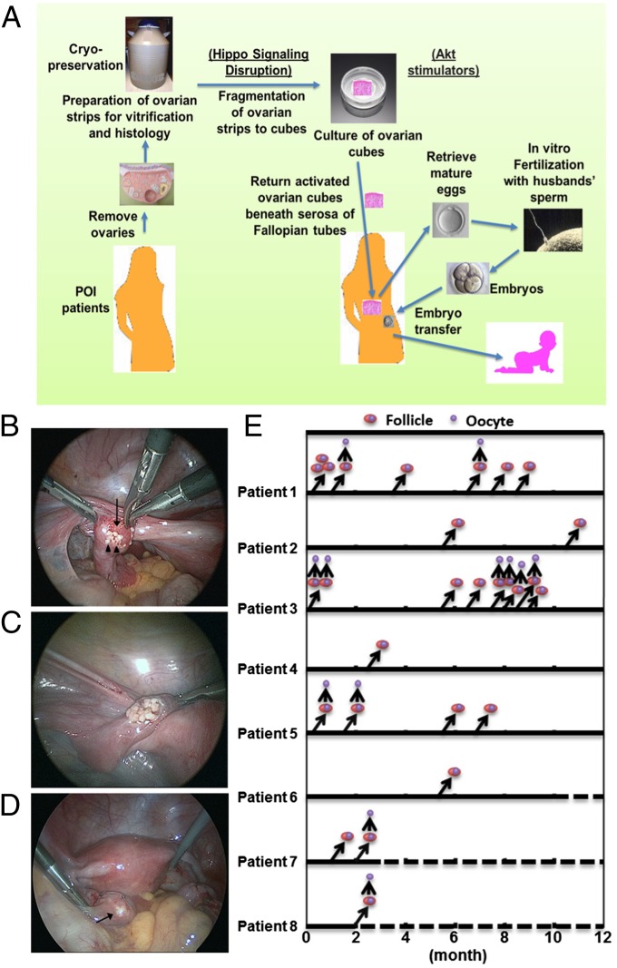
Fig. 4.
Ovarian fragmentation/Akt stimulation followed by autografting promoted follicle growth in POI patients to generate mature oocytes for IVF–embryo transfer, pregnancy, and delivery. (A) Under laparoscopic surgery, ovaries were removed and cut into strips. Ovarian strips from POI patients were vitrified. After thawing, strips were fragmented into 1–2 mm2 cubes, before treatment with Akt stimulators. Two days later, cubes were autografted under laparoscopic surgery beneath serosa of Fallopian tubes. Follicle growth was monitored via transvaginal ultrasound and serum estrogen levels. After detection of antral follicles, patients were treated with FSH followed by hCG when preovulatory follicles were found. Mature oocytes were then retrieved and fertilized with the husband’s sperm in vitro before cyropreservation of four-cell stage embryos. Patients then received hormonal treatments to prepare the endometrium for implantation followed by transferring of thawed embryos. (B) Transplantation of ovarian cubes beneath the serosa of Fallopian tubes. Arrow, fallopian tube; arrowheads, cubes. (C) Multiple cubes were put beneath serosa. (D) Serosa after grafting. Ovarian cubes are visible beneath serosa (arrow). (E) Detection of preovulatory follicles in grafts for oocyte retrieval. Following ultrasound monitoring, follicle growth was found in eight patients. After follicles reached the antral stage (>5 mm in diameter, right upward arrows), patients were treated with FSH followed by hCG for egg retrieval (upward arrows). Double circles represent preovulatory follicles, whereas single circles represent retrieved oocytes. Dashed lines depict ongoing observation.

