Fig. 6.
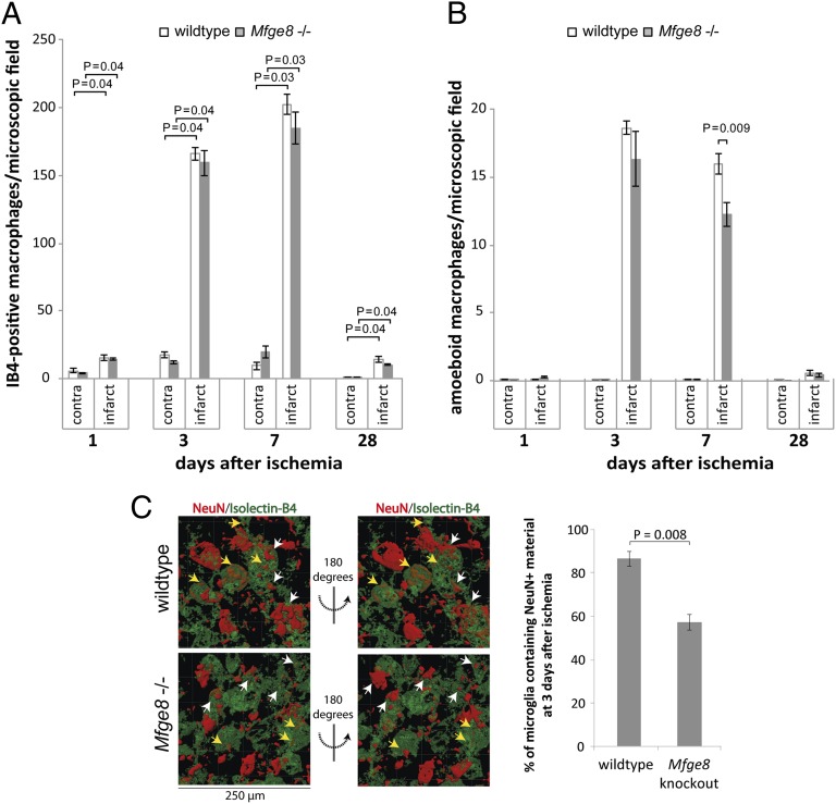
MFG-E8 deficiency inhibits phagocytosis of neurons after focal cortical ischemia. (A) A strong increase in the number of activated macrophages/microglia (isolectin-B4 stained) is observed at 3 and 7 d after ischemia, returns to baseline by 28 d, and does not differ between Mfge8 wild-type and knockout mice. (B) Densities of amoeboid macrophages/microglia, indicating phagocytic activity: Mfge8 knockout animals display decreased levels of phagocytic cells at day 7. (C) Quantification of 3D image reconstructions of confocal z-stacks of the infarct area. The number of microglia containing neuronal nuclear (NeuN+) material is significantly reduced in Mfge8 knockout animals at 3 d after ischemia (n ≥ 5 animals per condition). Yellow arrows indicate neuronal material scored as engulfed, white arrows indicate neurons that are not contained within microglia.

