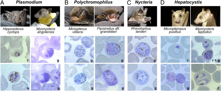
Fig. 3.
Hemosporidian parasites and their host species. Shown are captured bats and representative micrographs of Giemsa-stained thin blood films of their respective hemosporidian parasite blood stages (r, ring stage; s, schizont; g, gametocyte). (A) P. cyclopsi and P. voltaicum blood stages isolated from H. cyclops and M. angolensis, respectively. (B) Polychromophilus gametocytes isolated from miniopterid and vespertilionid bats. (C) Nycteria gametocytes isolated from two rhinolophid bats. (D) Hepatocystis blood stages isolated from six pteropodid bats. Shown are two (Micropteropus pusillus, Myonycteris leptodon) of six host species and different blood stages of their parasites. Micrographs were taken at 1,000× magnification.

