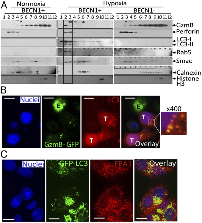
Fig. 3.
In vitro tumor cells degrade NK cell-derived granzyme B in lysosomes via autophagy. (A) NK cells were cocultured with autophagy-competent (BECN1+) or -defective (BECN1−) normoxic or hypoxic MCF-7 cells. Cell lysates of separated tumor cells were subjected to subcellular fractionation. Fractions 1 to 12 were characterized by Western blot using the indicated antibodies. * indicates an unspecific band. (B) Chloroquine-treated hypoxic MCF-7 cells (T) were cocultured with GzmB–GFP–expressing NK cells (E). Target cells were stained with an Alexa-Fluor-568–coupled rabbit anti-LC3 antibody to visualize autophagosomes (red) and with DAPI to stain nuclei (blue). Colocalization of GzmB with LC3 in autophagosomes was visualized by confocal microscopy using a 100× oil immersion objective. An enlarged image (400×) of the overlay (box) is shown. (Scale bar, 10 µm.) (C) Chloroquine treated GFP–LC3 –expressing MCF-7 cell were cultured under hypoxia stained with anti-EEA1 Alexa-Fluor-568–coupled anti-rabbit IgG antibody (red) and DAPI for nuclei. Cells were analyzed by confocal microscopy. The overlay image shows colocalization of LC3 and EEA1 (yellow dots). (Scale bar, 10 µm.)

