
Fig. 4.
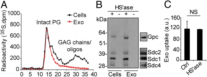
HSPGs are sorted to exosome vesicles but have no role in their uptake. (A) GBM cells were metabolically labeled with [35S]sulfate, and 35S-PGs and GAGs/oligosaccharides from cell and exosome lysates were size-fractionated on a Superose 6 column as described in SI Materials and Methods. (B) Isolated PGs from lysates of exosomes or GBM cells were untreated (−) or digested with heparinase III and ABC lyases (+). The digest products were separated by SDS page, and HSPG core proteins were visualized by immunoblotting with 3G10 anti-ΔHS antibody. Typical positions of members of the glypican and syndecan HSPG families are indicated (Right). (C) PKH-labeled exosomes (40 μg/mL) were untreated (Ctrl) or digested with heparinase I and III lyases (HS’ase), and then analyzed for uptake by GBM cells for 1 h by flow cytometry. Data shown represent the mean ± SD from three independent experiments, each performed in duplicate. NS, not significant (i.e., no statistically significant difference).