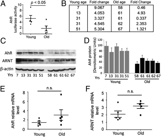
Fig. 2.
Endogenous AhR activity in human RPE cells decreases as a function of age. Each in vitro experiment was performed a minimum of three times. Data are means ± SEM for all graphs, P < 0.05. Representative Western blots from four independent experiments are shown. (A) Box plot of endogenous AhR activity in RPE cell lines derived from young (<55 y; n = 5) and old (>55 y; n = 5) human donors (n = 5 per age group) normalized to AhR activity in ARPE19 cells. Each statistical significance was analyzed by Student’s unpaired one-tailed t test; P < 0.05. (B) Details of AhR fold-activity changes and ages of each donor in young and old groups. (C) Western blot of AhR, ARNT, and housekeeping protein β-actin in young and old human donor RPE cell lines. (D) Densitometry measurements of the intensity of the AhR Western blot bands normalized to beta-actin for cell extracts. Box plot of (E) AhR and (F) ARNT mRNA relative to housekeeping gene 36B4 in young and old donor cell lines.

