Fig. 7.
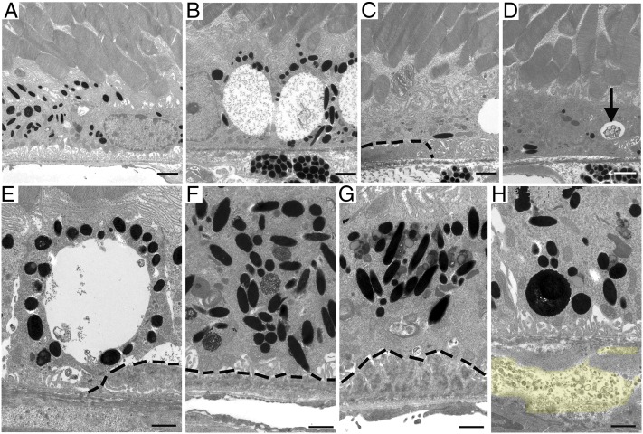
Ultrastructural changes in 16-mo-old AhR−/− mice. Electron micrographs of RPE/Bruch’s membrane/choroidal junction in (A) 16-mo-old wild-type mice appear normal whereas age-matched AhR−/− mice show (B) extensive vacuolization, (C) RPE atrophy overlying basal laminar deposit, (D) RPE atrophy, depigmentation, and vacuole containing membranous material (black arrow), (E) large vacuole adjacent basal laminar deposits, (F) hyperpigmentation, and accumulated lipofuscin in RPE cells overlying thin, and (G) thick continuous basal laminar deposits and (H) accumulation of basal linear like-deposit material (yellow highlight) within Bruch’s membrane. Basal laminar deposits are outlined by black dashed lines. n = 6–10 images per mouse, n = 11–12 mice per genotype were examined. (Scale bars: A–D, 1 μm; E–H, 2 μm.)

