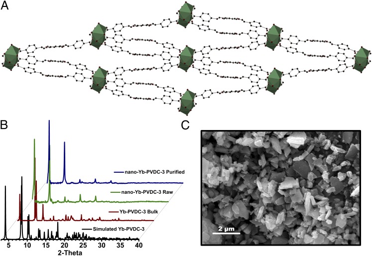
Fig. 1.
(A) Crystal structure of Yb-PVDC-3 viewed along the a-crystallographic axis. Yb3+ is shown as a polyhedron (C, dark gray; O, red; Yb3+, green). (B) PXRD patterns for simulated and bulk Yb-PVDC-3 and raw and purified nano-Yb-PVDC-3. (C) SEM image of nano-Yb-PVDC-3; average dimensions (±SD) are 0.5 (±0.3) μm (length), 316 (±156) nm (width), and 176 (±52) nm (thickness).

