Fig. 3.
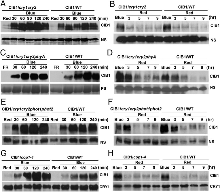
Lack of effect of CRY, phyA, and COP1 on CIB1 protein expression. (A, C, E, and G) Transgenic plants expressing the 35S::Myc–CIB1 transgene in wild-type (WT) or the indicated mutant backgrounds (cry1cry2, cry1cry2phyA, cry1cry2phot1phot2, or cop1) were grown in LD (16hL/8hD) for 3 wk and exposed to red light (20 μmol⋅m−2⋅s−1) for 16 h (A, E, and G) or exposed to far-red light (5 μmol⋅m−2⋅s−1) for 16 h (C) and then transferred to blue light (35 μmol⋅m−2⋅s−1) for the indicated time before sample harvest. (B, D, F, and H) Alternatively, the 3-wk-old plants were exposed to blue light (35 μmol⋅m−2⋅s−1) for 16 h and then transferred to red light (20 μmol⋅m−2⋅s−1) for the indicated time. Samples were fractionated by 10% SDS/PAGE, blotted, and probed by the anti-Myc antibody (CIB1). CRY1 or nonspecific bands (NS) are shown as the loading controls. Because of uncontrolled exposure times of ECL of different immunoblots, results of different blots are not directly comparable.

