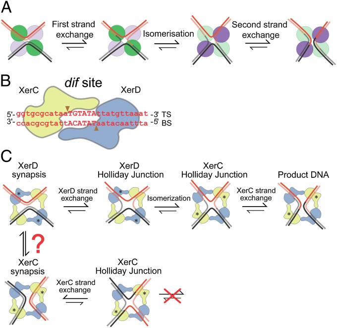
Fig. 1.
Tyrosine family site-specific recombination. (A) Schematic showing tyrosine recombinase pathway. The green and violet circles indicate recombinase protomers, with the color being highlighted when a given recombinase pair is active. Isomerization of the HJ intermediate plays an essential role in the reciprocal catalytic switches in recombinase activity. (B) XerCD–dif core site. XerC (yellow) and XerD (blue) bind to their respective DNA dif half-sites. TS and BS are the “top” and “bottom” strands, respectively. The scissile phosphates, indicated by the arrowheads, flank the central region, TGTATA. (C) XerCD–dif recombination at dif as envisaged using the Cre–loxP paradigm (6) (active monomers are labeled with asterisks). FtsK activated pathway is shown at the Top; an XerD-active synapsis (XerD*) leads to a XerD–HJ, which is subsequently isomerized to a XerC–HJ, which is resolved by XerC to recombinant products. In the absence of FtsK, XerCD–dif synaptic complexes (XerC*) can form and undergo cycles of XerC-mediated strand exchanges (Bottom). XerC and XerD synaptic complexes can only be interconverted by changing the path of the DNA helix in a complex, or by breaking the recombinase–recombinase interactions involved in synapsis and reforming the complementary synaptic complex de novo.

