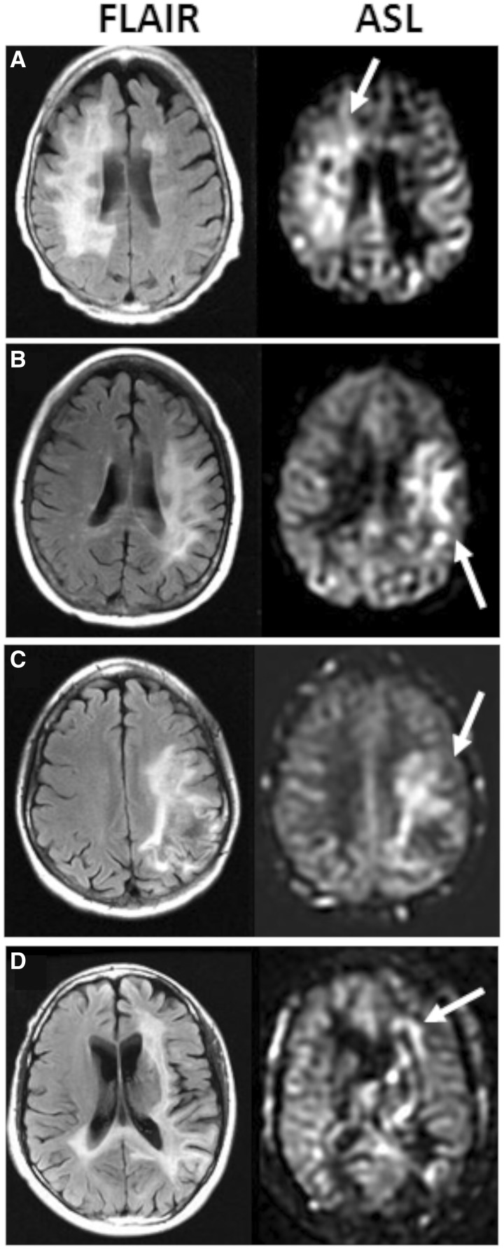
Figure 1.
PML lesions contain areas of elevated perfusion. Four representative cases of PML lesions on FLAIR with corresponding ASL perfusion sequences are shown. Areas of increased perfusion are marked by an arrow. (A) Right frontal lesion in a PML progressor. HLF of the PML lesion was 10.9%. (B) Left fronto-parietal lesion in a PML progressor with an HLF of 26.0%. (C) Left fronto-parietal lesion in a PML survivor with an HLF of 34.6% (D) Left fronto-parietal and splenium of corpus callosum lesion in a PML progressor with an HLF of 11.3%. Perfusion was more commonly elevated along the edge rather than the core of the PML lesions (B, C and D).

