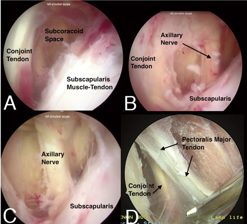
Figure 3.

A) The conjoint tendon and the subscapularis muscle tendon units define the anterior and posterior boundaries of the subcoracoid space, respectively, and can be visualized from a lateral viewing portal. B) With the arthroscope advanced more medially compared with Figure 3a, the deep aspect of the conjoint tendon and medial subscapularis muscle tendon unit can be seen. After a careful bursectomy in this area, the axillary nerve can also been seen along the lower border of the subscapularis. C) The axillary nerve visualized in the subcoracoid space from a lateral viewing portal. D) The conjoint tendon and the deep portion of the pectoralis major tendon can be visualized during the dissection required for an arthroscopic biceps tenodesis. This view is appreciated from a lateral viewing portal at the junction of the anterior and middle thirds of the acromion with the arm in a forward flexed and slightly abducted position.
