Figure 1.
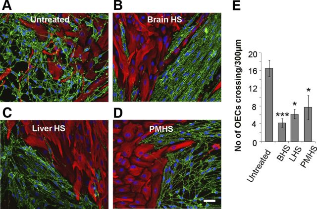
HS from a variety of tissue sources can induce a boundary in OEC–astrocyte confrontation assays. A–D, 30 μg/ml HS purified from brain (BHS, B), liver (LHS, C), and porcine intestinal mucosa (PMHS, D), were added to confrontation assays of OECs and astrocytes and compared with an untreated control (A). After 2 d of treatment, cells were fixed and stained for GFAP (red) and p75NTR (green). E, The numbers of OECs mingling with astrocytes across a 300 μm line were counted. HS from all three tissue sources prevented OECs from crossing into the astrocyte monolayer, resulting in the formation of a boundary. Error bars indicate ± SEM. Scale bar, 50 μm. *p < 0.05, ***p < 0.001 versus control.

