
Figure 2.
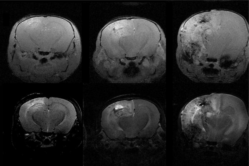
Representative trans-axial MR images of mice at two (left), three (middle) and four months (right) following fractionated hemispheric irradiation. The upper half of this panel are contrast-enhanced, T1-weighted, gradient-echo images; the lower half of the panel are T2-weighted spin-echo images. Consistent with other animal studies, the left side in each of these images corresponds with the left hemisphere of the mouse’s brain, which is the opposite of the convention used when displaying human images. Note the progressive increases in contrast enhancement and T2 signal hyper-intensity with increasing time following irradiation.