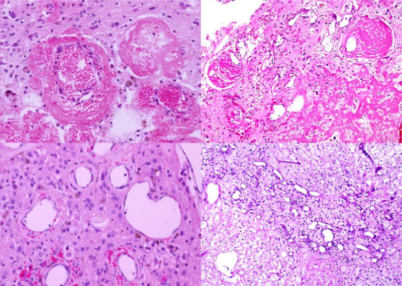
Figure 3.

Representative histological images from (left) a mouse three months after fractionated hemispheric irradiation and (right) a human subject with radiation necrosis. The histology illustrates changes typically observed in radiation necrosis, including (top) fibrinoid vascular necrosis and (bottom) vascular telangiectasia. The similarities between the mouse and human histology are evident by comparing the left and right panels of this figure.
