Figure.

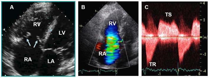
Characteristic echocardiographic images from a patient with carcinoid heart disease. Panel A. Apical 4-chamber view showing marked right atrial (RA) and right ventricular (RV) enlargement. The tricuspid leaflets (arrows) are thickened, retracted and fixed leading to both tricuspid regurgitation (TR) and tricuspid stenosis (TS). Panel B. Apical 4-chamber view showing severe TR by color flow Doppler. Panel C. Continuous-wave Doppler showing both TS and TR. On the TR jet notice the classic dagger-shaped Doppler spectral profile with an early peak pressure followed by a rapid decline, indicative of markedly increased RA pressure.
