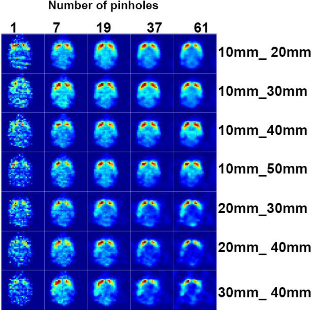
Figure 10.
Center axial slice of the mouse brain phantom reconstructed with different combinations of front and back detector distances to the collimator (10mm_20mm, 10mm_30mm, 10mm_40mm, 10mm_50mm, 20mm_30mm, 20mm_40mm, 30mm_40mm) and number of pinholes (1, 7, 19, 37, 61). The geometry for the acquisition is illustrated in figure 4 (top). The left side of the brain is sampled at a closer distance to the collimator.

