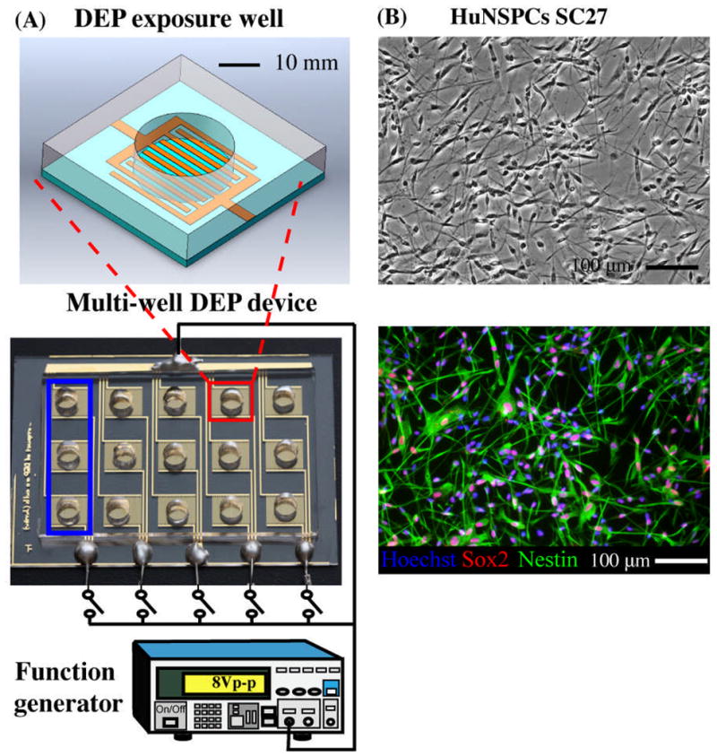
Fig. 1. Multi-well device and experimental setup for testing effects of DEP force on NSPCs.

(A) DEP exposure well (top schematic) contains interdigitated gold-plated electrodes at the bottom of the well (sides of well are PDMS). Fifteen individual wells (red square) make up the multi-well DEP device (bottom schematic), which is connected to a function generator to energize the electrodes (energized by 8Vpeak-peak). The bottom electrical head serves as a DEP force switch for each group of three exposure wells (blue square). The symbol,
 , represents a switch in the circuit. (B) Undifferentiated HuNSPCs (SC27) are grown as adherent cultures (phase contrast panel) and express the neural stem cell markers Sox2 and Nestin (fluorescence panel, 98.0 +/− 0.5% of the cells express either Sox2 or Nestin and 78.6 +/− 2.7% co-express both markers, N=1300 cells). All cell nuclei are stained with Hoechst. NSPCs are grown as adherent cultures but were dissociated to form a cell suspension for all DEP exposure experiments. Data are represented as mean ± SE.
