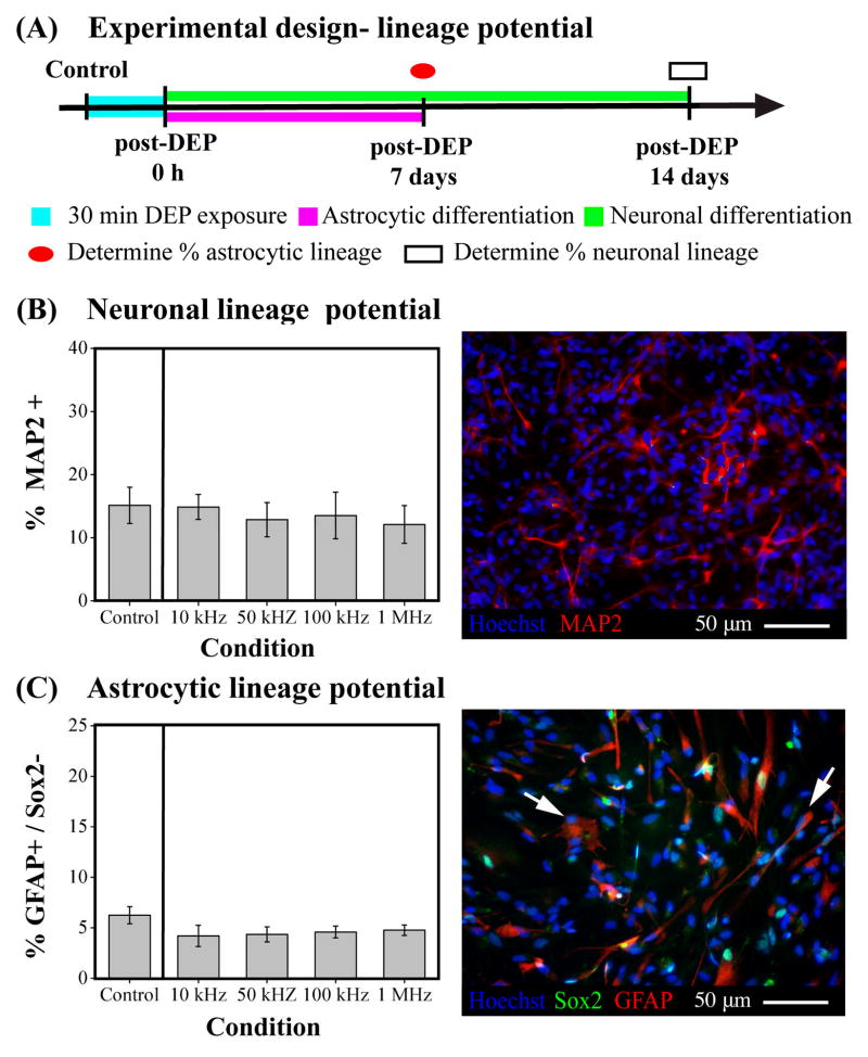
Fig. 5. DEP exposure does not interfere with differentiation of NSPCs into neuronal and astrocytic lineages.
(A) Schematic portrays the experimental design to test effects of DEP on neuronal and astrocytic lineage potential of NSPCs. (B) Left panel: The neuronal lineage potential of SC27 HuNSPCs (as shown by generation of MAP2+ cells) is not altered by long-term DEP exposure at various DEP frequencies. Control cells were not exposed to DEP force. Right panel: Image shows HuNSPCs (SC27) differentiated for 14 days and stained with MAP2 antibody (red) to detect neurons. All nuclei are stained blue. (C) Left panel: The astrocytic lineage potential of SC27 HuNSPCs (as shown by generation of GFAP+/Sox2− cells) is not modified by long-term DEP exposure at various DEP frequencies. Right panel: Image shows HuNSPCs (SC27) differentiated for 7 days and co-stained with GFAP (red) and Sox2 (green) antibodies (GFAP+/Sox2− astrocytes are shown by arrows). All nuclei are stained blue. Data are represented as mean ± SE.

