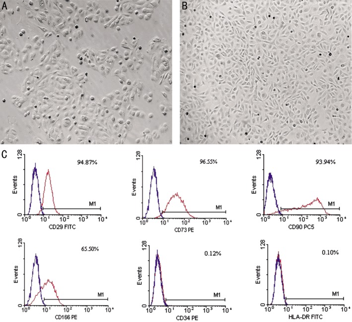
Figure 1. The morphology and phenotypical characterization of hAECs.
A: Cultured hAECs 48h after plating; B: Cultured hAECs confluence at day 7; C: Flow cytometry analysis of confluent hAECs. The results showed that the expression of stem cell markers CD29, CD73, CD90, CD166 was positive, the hemotopoietic marker CD34 was negative, and the expression of HLA-DR was absent. Magnification: 50×(A, B).

