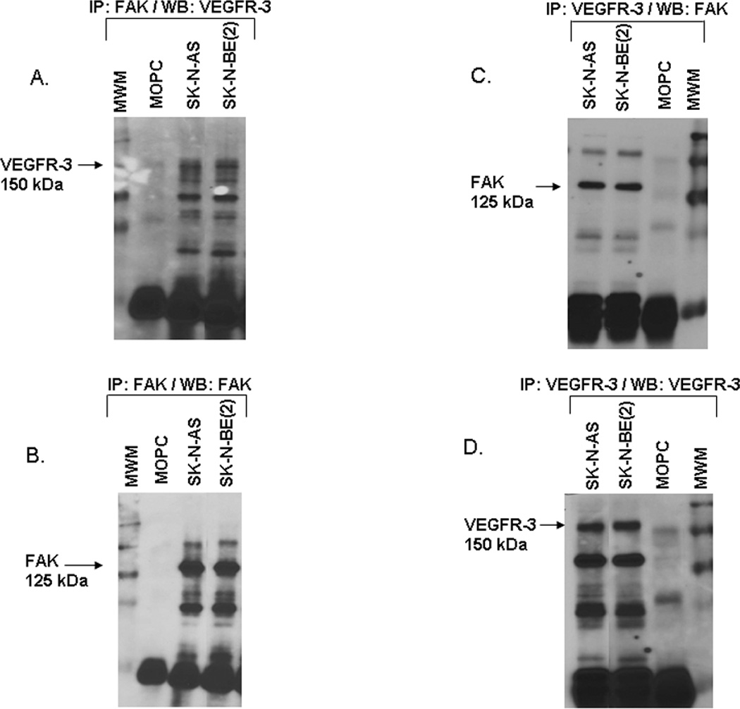
Figure 5. Interaction of VEGFR-3 and FAK in neuroblastoma cell lines.

A. Immunoprecipitation followed by Western blotting was used to detect FAK and VEGFR-3 interaction in the neuroblastoma cell lines. Immunoprecipitation with FAK antibody followed by Western blotting for VEGFR-3 showed a band for VEGFR-3 at the expected 150 kDa in both cell lines. B. Western blotting of that same membrane for FAK, revealed a FAK band present at 125 kDa. C. Immunoprecipitation with VEGFR-3 antibody followed by Western blotting for FAK showed a band for FAK at the expected 125 kDa in both cell lines. D. Western blotting of the same membrane for VEGFR-3, detected a band at the expected 150 kDa. These data showed that FAK and VEGFR-3 interact in both neuroblastoma cell lines.
