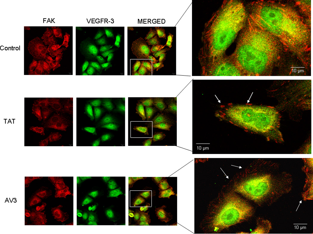
Figure 7. AV3 peptide treatment did not result in a loss of FAK from the focal adhesions in the SK-N-AS cell line.

Immunofluorescence staining followed by confocal microscopy was employed to evaluate FAK and VEGFR-3 in the SK-N-AS cell line after AV3 treatment for 24 hours. In these cells, AV3 did not lead to a loss of FAK from the focal adhesions (white arrows bottom fourth panel), and did not affect the dual staining of VEGFR-3 and FAK (bottom third and fourth panels). The control peptide (TAT) had no effect upon FAK at the focal adhesions (white arrows middle fourth panel).
