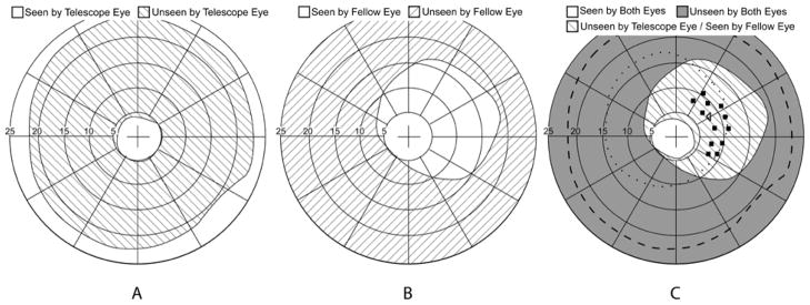
Figure 3.

An example of binocular visual field plots obtained for a right strabismic patient wearing a 3x Galilean bioptic on the left eye. (A) Monocular viewing through the telescope without shutter lenses (fellow-eye patched) showing the ring scotoma of the telescope. (B) Asymmetric field of view through the fellow (right) eye shutter lens. Fixation and background were seen with both eyes while the perimetry stimulus was presented only to the fellow (non-telescope) eye. (C) Binocular viewing with the shutter lenses; fixation and background were seen by both eyes. The square symbols show the position of static stimuli (within the area of the telescope eye ring scotoma) presented to the temporal field of the fellow eye only. The left-facing triangle shows the position of the fixation catch trial static stimulus (in the ring scotoma) presented to the telescope eye only. The dashed line shows the outer boundary of the ring scotoma as measured in (A). The dotted line indicates the restriction of the telescope-eye shutter lens.
