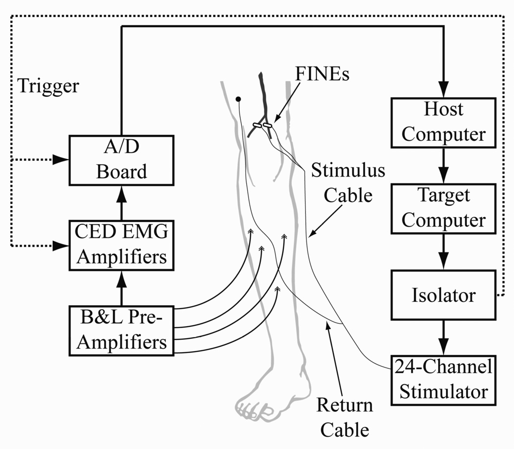
Figure 3.
Experimental setup for testing the FINE on the tibial or common peroneal nerve. A custom, current-controlled stimulator delivered stimulus pulses to the FINE. Differential EMG was collected from each of four muscles innervated by the target nerve. EMG was referenced to a ground patch (not shown), amplified, filtered, and collected.

