Fig. 5.

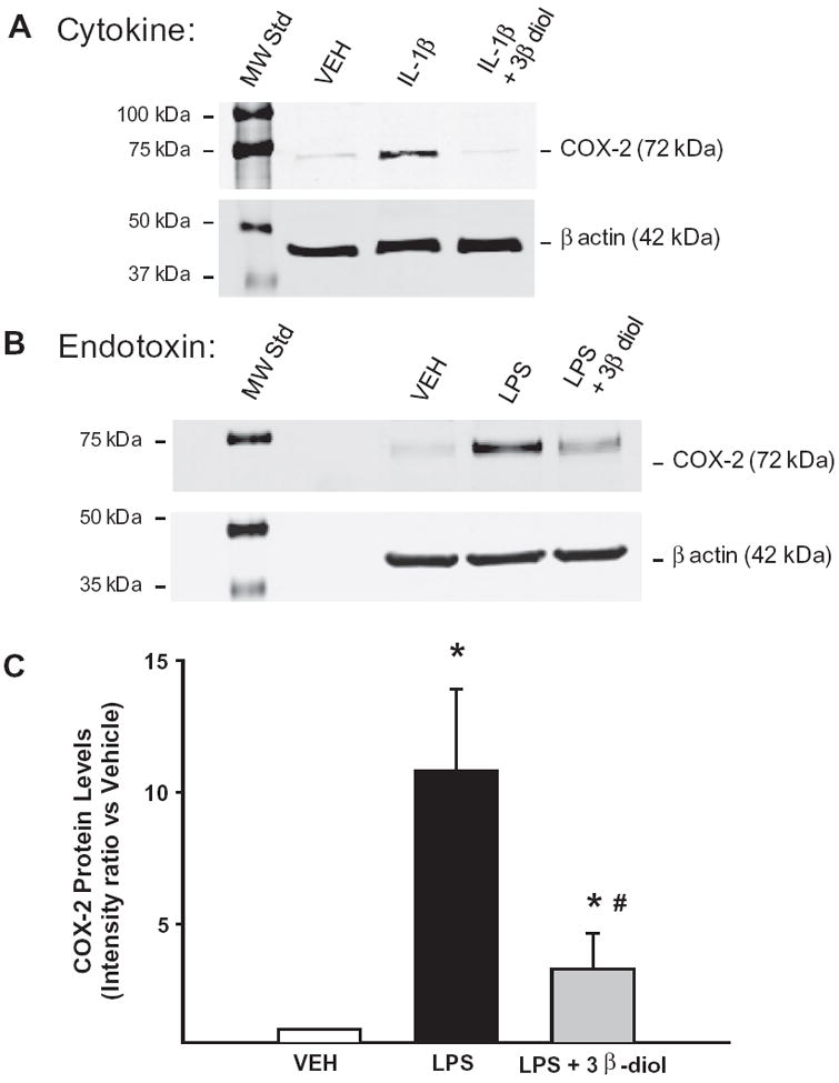
3β-Diol inhibited cytokine- and endotoxin-induced increases in COX-2 in rat mesenteric arteries. Cyclooxygenase-2 (COX-2) protein levels were assessed in isolated mesenteric arteries treated ex vivo with either cytokine or endotoxin in the absence or presence of 3β-diol. Panel A illustrates the Western blot for COX-2 expression for the cytokine experiment: vehicle (VEH), interleukin-1 beta (IL-1β; 10 ng/ml), or IL-1β (10 ng/ml) plus 3β-diol (10 nM). Panel B illustrates the LPS experiment: VEH, the endotoxin lipopolysaccharide (LPS; 100 μg/ml), or LPS + 3β-diol. β Actin served as a loading control. Panel C: Graph showing COX-2 levels after LPS treatment. *P < 0.05 vs. VEH, #P < 0.05 vs. LPS (n = 3).
