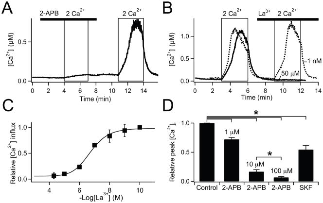
Fig. 4.
Pharmacological block of SOC in PDEC. Single-cell Ca2+-photometry shown in (A) and (B), and summarized data shown in (C) and (D). (A) Cells were pretreated with 5 μM thapsigargin for 5 min to deplete the Ca2+ stores in Ca2+-free Ringer’s. Ca2+ influx through SOC, upon the switch to 2 mM external Ca2+, was blocked by 100 μM 2-APB (bar) (n = 6, (A)) and 50 μM La3+ (bar) (n = 3, solid line in (B)), but not, 1 nM La3+ (n = 3, dotted line in (B)). (C) Dose response curve for La3+-mediated block of Ca2+ influx (IC50 = 199 nM, n = 3–7 for each concentration). (D) Block of Ca2+ influx by SOC blockers, 1, 10, 100 μM 2-APB (n = 11, 8, 6), and 50 μM SKF-96365 (n = 3). Peak [Ca2+]i values achieved by 2 mM external Ca2+ in the presence of blockers compared to control. The same experimental protocol was used as for (A). P* <0.05.

