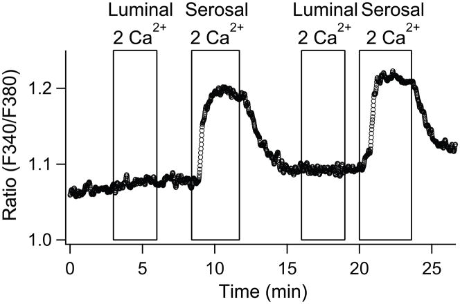
Fig. 5.
Localization of SOCs to the basolateral side of polarized PDEC. Representative of 4 experiments. A differentiated PDEC monolayer were pretreated with 5 μM thapsigargin for 5 min in Ca2+-free Ringer’s. External 2 mM Ca2+-containing solution was applied to either luminal/apical or serosal/basolateral sides of PDEC. [Ca2+]i is expressed as the fluorescence ratio at 340 and 380 nm.

