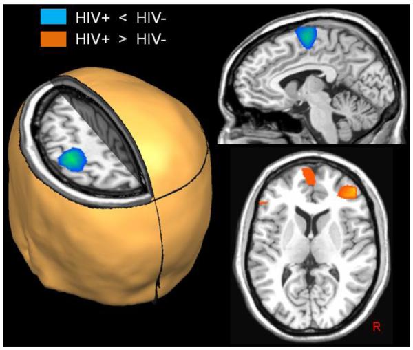
Figure 3.
Group Differences in Beta ERD Activity. Uninfected controls exhibited significantly stronger responses compared with HIV-infected patients (blue areas; p < 0.01, cluster-corrected) in the left precentral gyrus, SMA, and a small area of the right precentral gyrus (not shown). In contrast, significantly greater beta ERD responses were detected in the right dorsolateral prefrontal cortices (DLPFC), medial prefrontal cortices, and the left inferior frontal gyrus of HIV-infected patients relative to controls (orange areas; p < 0.01, cluster-corrected). All images are shown in neurological convention.

