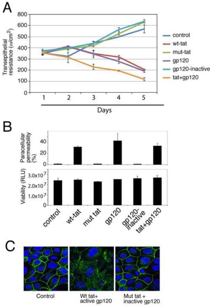
Fig. 2.
Role of HIV proteins tat and gp120 in the disruption of oral epithelial tight junctions. (A) Polarized oral epithelial cells were treated with an active and inactive recombinant HIV tat or gp120, separately or in combination. TER was measured daily. (B) (upper panel) The same cells were used to measure paracellular permeability after 5 days of treatment. IgG leakage into lower chambers is presented as % of apical value of tracer. (Lower panel) After measuring paracellular permeability, cells were examined for viability using an MTT assay. RLU, relative luminescence units. (C) At 5 days, untreated control cells and cells treated with inactive and active tat and gp120 were immunostained for occludin (green). Cell nuclei are stained in blue. (A and B) Error bars show ± s.e.m. (n=3).

