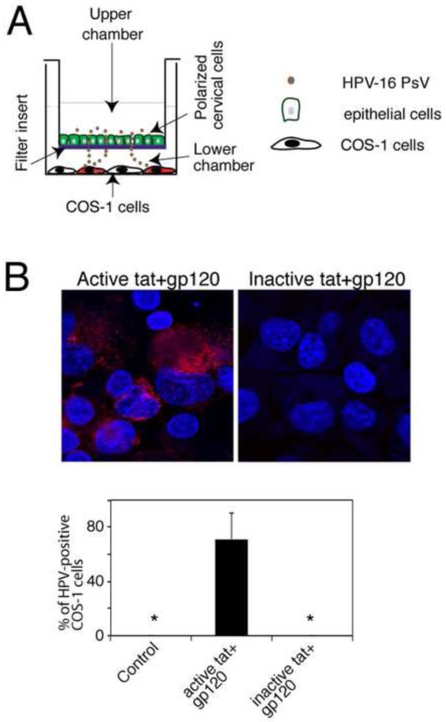
Fig. 3.
Role of tat- and gp120- disrupted epithelial TJ in paracellular spread HPV-16 PsVs. (A) Model of HPV-16 PsV paracellular passage between disrupted mucosal epithelia. Polarized cells were grown in the upper chambers of two-chamber Transwell filter inserts. COS-1 cells were grown in the lower chambers of the filter inserts, in the well where the filter inserts were then placed. To examine paracellular passage of HPV-16 PsVs, PsVs were added to the apical surfaces of polarized epithelial cells. At 2 h post-inoculation, filter inserts were removed, and the extent of paracellular passage of PsVs was determined in the COS-1 cells by detecting RFP fluorescence. (B, upper panel) Polarized oral epithelial cells were incubated for 5 days with HIV-tat and gp120 or their inactive forms. HPV-16 PsVs were then added to the apical surfaces and paracellular passage of HPV-16 PsVs across disrupted oral epithelium was examined by detection of RFP-positive COS-1 cells. (B, lower panel) Paracellular passage of HPV-16 PsVs across disrupted oral epithelium was quantitated by counting RFP-positive COS-1 cells. Results are presented as a % of RFP-positive cells. Error bars show ± s.e.m. (n=3). *, not detected.

