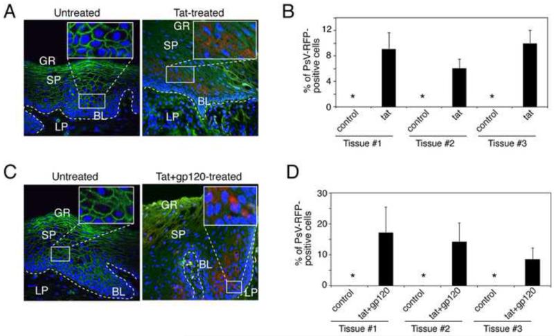
Fig. 4.
HIV-associated disruption of mucosal epithelium facilitates PsV entry into basal and parabasal cells. (A and B) Buccal explants were treated with HIV tat alone (A) or HIV tat and gp120 in combination (B) for 2 days and then exposed to HPV-16 PsVs for the next 3 days. Tissues were immunostained for ZO-1 (red) and analyzed for PsV-RFP by confocal microscopy. GR, granulosum; SP, spinosum; BL, basal; LP, lamina propria. (C and D) PsV penetration of HIV tat- and gp120-treated explants was quantified by counting RFP-positive epithelial cells; data are presented as the percentage of cells positive for PsV-RFP. Error bars show [g940] s.e.m. (n=10). *, not detected.

