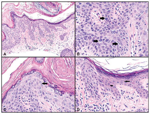
Fig. 1.
A–D) Bowen’s disease. A) Bowen’s disease on left and adjacent normal epidermis on right. Hematoxylin/eosin (H&E)-stained sections, magnification ×200. B) Multiple hyperchromatic cells in all layers of the epidermis, with crowding and irregular nuclei (sharply angulated pentagon shaped and curved cigar shaped – right-sided arrows). H&E sections, magnification ×600, cropped image. C) Another high-power field from same case showing cell with prominent nucleolus (arrow), which was visible at lower magnification. Irregular nuclei and crowding are also seen in this field. H&E sections, magnification ×600. D) Junction between Bowen’s disease and adjacent uninvolved epidermis for comparison. Cells in the uninvolved portion are round or oval in shape, without hyperchromasia and with delicate small nucleoli (arrows). In comparison, cells in the area of Bowen’s disease show irregular angulated shapes, hyperchromasia, increased nuclear/cytoplasmic ratio and crowding. H&E sections, magnification ×600.

