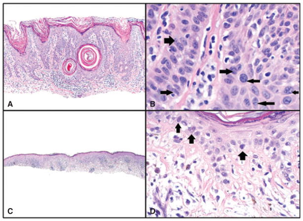
Fig. 3.
A and B) Seborrheic keratosis with inflammation. A) Hematoxylin/eosin (H&E) sections, magnification ×100. B) Nuclear crowding (right-sided arrows) and a clumped chromatin pattern (left-sided arrows) are present in areas of this benign lesion. Although some nuclei are compressed and elongated (left lower arrow), they did not meet our criteria for irregular nuclei, which require sharp angulations or curves in the nuclear membrane. Magnification ×600, cropped image. C and D) Benign lichenoid keratosis (lichen planus-like keratosis). C) H&E sections, magnification ×40. D) Hyperchromatic cells at the basal layer of the epidermis. Such cells were found focally in lesions with inflammation; however, their frequency and expression were much lower than in malignant lesions. Also, no foci show crowding, high nuclear/cytoplasmic ratio or irregular nuclei. H&E sections, magnification ×200.

