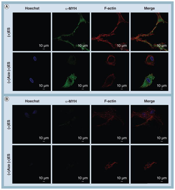
Figure 7. Confocal imaging of human mesenchymal stem cell protein localization and cell morphology following electrical stimulation.
(A) In the presence of ES, human mesenchymal stem cells cultured on poly(ε-caprolactone) substrates increased cell–cell contacts with neighboring cells and maintained strong expression of α-MYH as well as colocalization with F-actin. (B) In contrast, ES completely silenced expression of α-MYH for human mesenchymal stem cells on poly(ε-caprolactone) carbon nanotube substrates and caused cells to lose their elongated morphology. For cells cultured on either substrate, cotreatment with ES and Aza resulted in a complete deregulation of normal cell function as indicated by changes in cell morphology and either loss of α-MYH expression or colocalization with actin.
α-MYH: α-myosin heavy chain; Aza: Azacytidine; ES: Electrical stimulation; F-actin: Filamentous actin.

