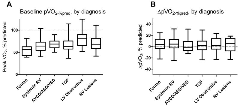
Figure 1. Baseline % predicted peak VO2(A) and change in % predicted peak VO2 between baseline and follow-up cardiopulmonary exercise tests(B) by congenital heart disease diagnostic category.
While baseline pVO2-%pred differed by diagnosis (Kruskal-Wallis, p=0.006), there was no such difference in ΔpVO2-%pred between diagnoses (Kruskal-Wallis, p=0.96). RV=right ventricular lesion, LV=left ventricular lesion, AVCD=atrioventricular canal defect, ASD/VSD=atrial/ventricular septal defect, TOF=tetralogy of Fallot

