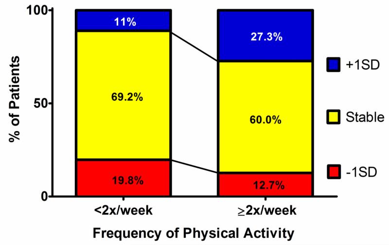
Figure 3. Proportion of patients having >1SD(±11%) change in peak VO2-abs between exercise tests.
Most patients had stable pVO2, independent of PA frequency. However, approximately twice as many patients in the lower exercise frequency group had a >1SD decrease in pVO2 compared with a >1SD increase. Conversely, among those engaging in frequent PA, more than twice as many improved their pVO2 >1SD compared to the number that had equivalently decreased pVO2.

