
Figure 7. Effects of Reelin fragments on APPsα in HEK293 cells.
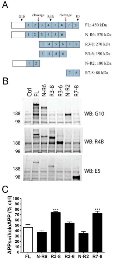
Schematic of Reelin fragments generated from proteolytic processing. Reelin repeat domains are numbered in blue. Red arrows represent cleavage sites. Black arrowheads represent antibody epitopes. (B) Western blots of cell lysates showing expression of Reelin fragments transfected into HEK293 cells. (C) ELISA quantification of APPsα levels in HEK293 cells co-transfected with APP751 (human, wild-type) and either full-length Reelin or individual Reelin fragments. Error bars represent s.e.m.; ***p<.001, relative to full-length Reelin (FL)