
Fig. 3.
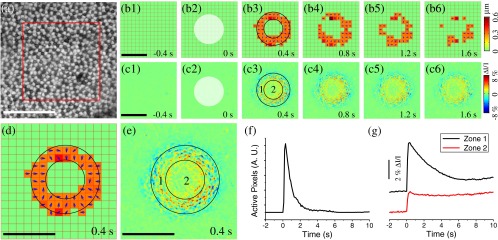
Photoreceptor displacements and intrinsic optical signal (IOS) responses stimulated by circular stimulus (in transverse plane) with a Gaussian profile (in axial plane). (a) NIR image of frog photoreceptor mosaic. Red rectangle indicated the area shown in Video 3 (QuickTime, 7.9 MB) [URL: http://dx.doi.org/10.1117/1.JBO.18.10.106013.3] which displays a pair of pre- and poststimulus images alternating repeatedly 20 times. (b) Localized retinal displacements associated with circular stimulus. This stimulus had a Gaussian profile in axial plane [Fig. 1(b)]. The same methods as those in Figs. 2(e) and 2(f) were used here to produce the displacement maps. (c) IOS maps. reflected the light intensity change and was the background light intensity. Zone 1 corresponds to the area within the inner ring. Zone 2 corresponds to the annular area. The stimulus was delivered at time 0. (d) Enlarged view of b3. (e) Enlarged view of c3. Scale bars indicate 100 μm. (f) Dynamic change of the number of active pixels in (b). The pixel with nonzero value was defined as active. (g) Temporal IOS profiles.
Click here for additional data file. (7.7MB, mov)