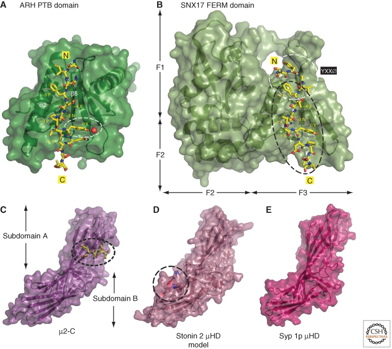
Figure 4.
PTB domains and μHDs in cargo signal recognition. (A) Combined ribbon and space-filling surface representation of the ARH PTB domain bound to the LDL receptor SINFDNPVYQKT sorting-signal peptide shown in stick representation (PDB ID: 3SO6) (Dvir et al. 2012). The location of a water molecule hydrogen bonded to the position 0 tyrosine is indicated by a red sphere within the marked PTB-domain tyrosine-binding site. (B) Combined ribbon and surface representation of the SNX17 FERM-like domain complexed with the TYGVFTNAAYDPT signal from P-selection (PDB ID: 4GXB) (Ghai et al. 2013). Shown in the same relative orientation as A with the positioning of the F1–F3 subdomains indicated and the YXXØ and [FY]XNPX[YF] interaction surfaces highlighted. (C–E) Combined ribbon and surface representations of the AP-2 µ2 subunit bound to the YQRL signal from TGN38 (PDB ID:1BXX) (Owen and Evans 1998), a homology model of the human Stonin 2 mHD generated by Phyre2 (Kelley and Sternberg 2009), and the Saccharomyces cerevisiae Syp1p μHD (PDB ID: 3G9H) (Reider et al. 2009). The relative positioning of the YXXØ interaction surface in subdomain A on μ2, and the biochemically defined synaptotagmin 1 C2A-binding site on subdomain B of stonin 2 (Jung et al. 2007) are indicated. Three side chains on the stonin 2 µHD (Lys783, Tyr784, Glu785) implicated in C2A binding are highlighted.

