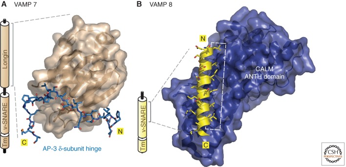
Figure 5.
Decoding folded endocytic sorting signals. (A) Schematic representation of the overall architecture of VAMP 7 with the amino-terminal longin domain, the vesicle-SNARE (v-SNARE) helical segment, and the transmembrane (Tm) domain indicated. A combined ribbon and surface representation of the globular VAMP 7 longin-domain “signal” bound by the extended AP-3 δ-subunit hinge polypeptide (blue; in stick representation) is also shown (PDB ID: 4AFI) (Kent et al. 2012). Notice the insertion of several hydrophobic side chains from the δ hinge into complementary acceptor cavities arrayed around the regulatory longin-domain circumference. (B) Schematic of the architecture of VAMP 8, which lacks the autoinhibitory longin domain. A representation of the amino-terminal portion (residues 15–38) of the α-helical v-SNARE domain of VAMP 8 complexed with the α-solenoid folded ANTH domain from CALM (PDB ID: 3ZYM) (Miller et al. 2011). The structured VAMP 8 amphipathic α helix orients seven aliphatic side chains to insert into a vertical groove along the ANTH surface (white dashed line).

