
Figure 1.
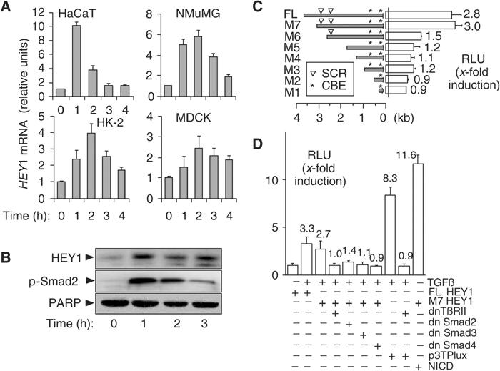
HEY1 is a direct target gene of TGF-β/Smad signalling. (A) Bar graphs show averages of relative HEY1 mRNA abundance determined by quantitative RT-PCR analysis in keratinocytes (HaCaT), mouse mammary gland epithelial cells (NMuMG), human kidney proximal tubular epithelial cells (HK-2), and canine kidney distal tubular epithelial cell line (MDCK). (B) Immunoblots show HEY1 and C-terminally phosphorylated Smad2 p-Smad2 proteins in keratinocytes stimulated with TGF-β. PARP, loading control. (C) Bars show average (N=3) fold induction by TGF-β of relative luciferase units (RLUs) in keratinocytes transfected with reporter constructs of human HEY1 promoter deletions (full-length FL, M7 to M1). (▿) positions of SCRs; (*) positions of NICD-responsive CBEs. (D) Bars indicate average (N=3) fold induction by TGF-β of luciferase activity (RLU) in keratinocytes cotransfected with luciferase reporter constructs (FL-HEY1, M7 HEY1, or 3TPLux) and plasmids expressing dominant-negative mutants of type II TGF-β receptor (dnTbRII), Smad2, 3 and 4 (dn Smad2, 3 and 4), or NICD. p3TPlux, positive control for TGF-β/Smad; NICD, positive control for activation of CBE sites.