
Figure 4.
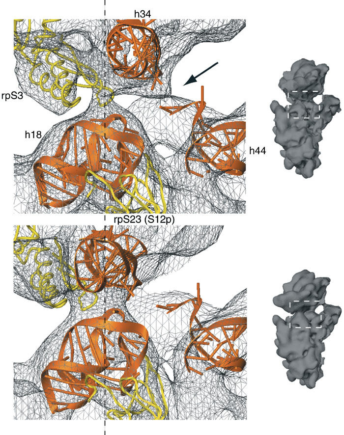
Rearrangement of the latch region of the 40S subunit. Comparison of the latch region in the 80S·eEF2·sordarin complex (upper panel) with the POST 80S ribosome (lower panel) (Spahn et al, 2001a). The corresponding cryo-EM densities are shown as a gray wire-mesh. Docked models for h18, h34 and the top of h44 are shown as orange ribbons, and models for rpS3 and rpS23 (S12p) as yellow ribbons. The dashed line goes through the center of the latch. Small inset on the right: the 40S subunits as an orientation aid. The white, dashed box indicates the latch region. The arrow in the upper panel highlights an additional connection between the head and the body of the 40S subunit that is present in the 80S·eEF2·sordarin complex.