
Figure 3.
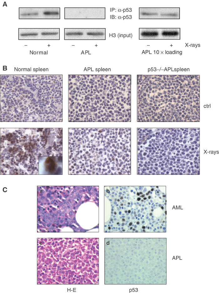
APL blasts have low levels of p53 and show no stabilization of p53 after X-ray treatment. (A) Normal and leukemic mice were given whole-body irradiation (9 Gy) or left untreated as indicated. The animals were killed 6 h later, spleen cells were isolated and nuclear extracts were prepared. The analysis was performed by immunoprecipitation (IP) followed by immunoblotting (IB) as indicated. Inputs were normalized according to histone H3 levels verified by immunoblotting prior to IP. Note that the starting material for the IP from leukemic samples was 10-fold that of normal samples due to the low expression of p53 in leukemic blasts. (B) Frozen spleen sections of normal and leukemic animals treated with X rays (or untreated mice as control) were stained with an anti-p53 antibody. An immature, wild-type myeloid precursor (by morphological criteria) showing strong p53 inducibility upon X-ray treatment is shown at a higher magnification in the inset. P53−/− spleens are shown as a negative control for p53 expression. (C) Immunohistochemistry analysis of p53 levels in human APL and non-APL samples. A representative case of AML (M2), diffusely immunoreactive for p53, is shown in the upper part of the panel. A case of APL, unreactive for p53, is shown in the lower part of the panel (40 × original magnification). (a and c) Hematoxylin and eosin, (b and d), hematoxylin counterstain.