
Figure 5.
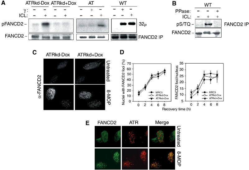
ATR is involved in ICL-dependent FANCD2 phosphorylation. (A) In vivo phosphoradiolabeled FANCD2 protein was immunoprecipitated from WT (HeLa), A-T derived, ATRkd-Dox or ATRkd-expressing (ATRkd+Dox) cells in response to 8-MOP (10 μM)+UVA (10 kJ/m2) or 20 Gy of IR. The FANCD2 immunoprecipitates were prepared 3 h after treatment and analyzed by SDS–PAGE and autoradiography. Western blotting with anti-FANCD2 antibodies is reported to show equal loading of the proteins into the gel. (B) FANCD2 was immunoprecipitated from ICL-treated HeLa cells and analyzed by an anti-pS/TQ specific antibody. (C, D) FANCD2 foci formation and quantitation in WT and ATR inactivated cells after ICL treatment (8-MOP (10 μM)+UVA (10 kJ/m2)) as observed by immunostaining of the cells 6 h after treatment with an anti-FANCD2 antibody. (C) Formation and colocalization of FANCD2 and ATR foci in response to 8-MOP (10 μM)+UVA (10 kJ/m2) exposure analyzed by immunostaining of cells with an anti-FANCD2 antibody (green) and an anti-ATR antibody (red). A representative nucleus obtained from cells fixed before and 6 h after treatment is displayed.