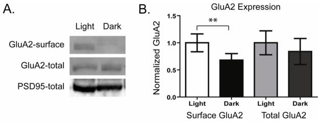
Figure 1. Darkness decreases surface GluA2 levels in retinal neurons.

A. Surface proteins were labeled with biotin in retina lysates isolated from rats housed either in light (Light) or darkness (Dark) for the preceding 12 hours. Surface proteins isolated by avidin-bead precipitation were separated by SDS-PAGE. Western blots were probed to detect surface as well as total GluA2 AMPAR levels. β-Actin or PSD95 was probed to control for loading differences. B. Quantitation of experiments as in A. Surface and total GluA2 labeling intensities are normalized to mean values of the light exposed samples. Results show that light deprivation reduces surface but not total GluA2 levels in retinal neurons (n=6, ** p<0.01).
