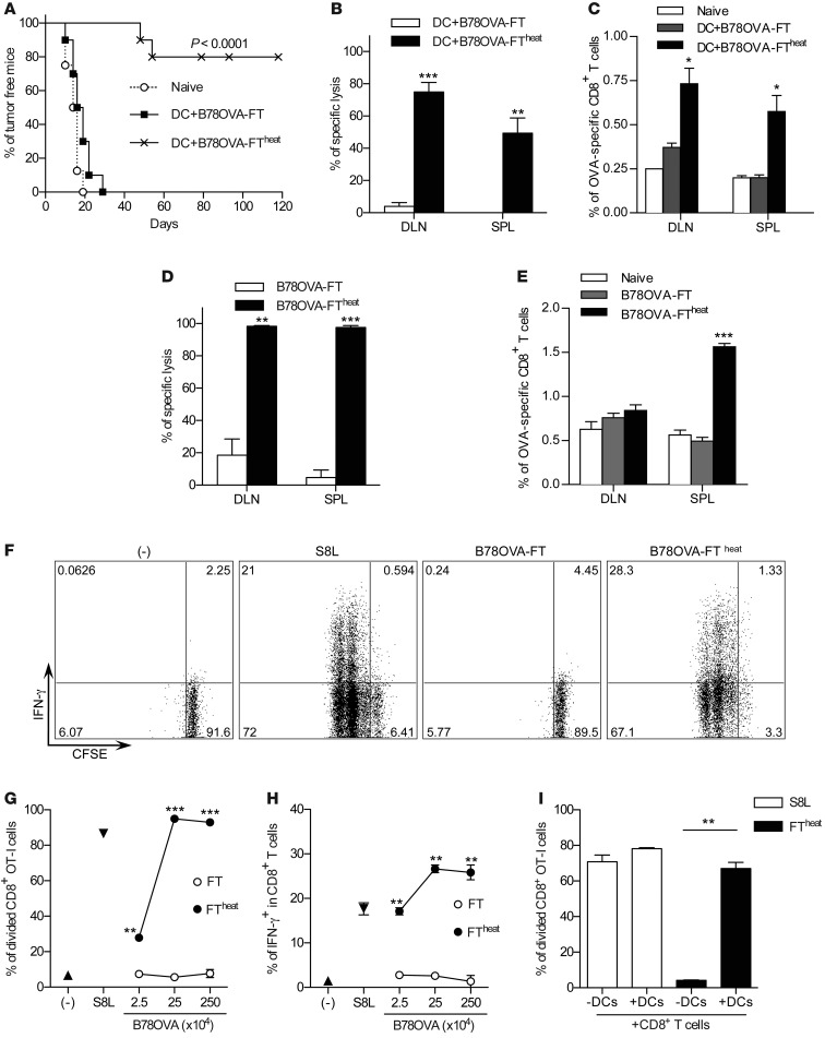
Figure 1. Primary FT necrotic cells do not induce CD8+ T cell activation.
(A) On days 0 and 2, splenic CD11c+ DCs were pulsed with SF of B78OVA-FT or B78OVA-FTheat cells. DCs were washed and injected into mice. On day 7, recipients were challenged with live B16OVA, and tumor growth was monitored. Data are from 2 independent experiments (n = 8–10 per group). P < 0.0001, log-rank (Mantel-Cox) test. (B and C) Mice were vaccinated as in A, and OVA-specific lysis (B) and percent OVA-specific CD8+ T cells (C) in draining LNs (DLN) and spleens (SPL) were determined on day 7. Data are from 1 representative of 2 experiments (n = 3 per group). (D and E) Mice were vaccinated with 5 × 10⁶ B78OVA-FT or B78OVA-FTheat cells on days 0 and 2. On day 7, OVA-specific lysis (D) and percent tetramer-positive cells (E) was determined. Data are representative of 3 experiments (n = 3 per group). (F–H) SF from B78OVA-FT was incubated at 70°C (FTheat) and cultured in vitro with OT-I splenocytes. Proliferation and IFN-γ expression of CD8+ T cells was analyzed after 48 hours. (F) IFN-γ release by proliferating CD8+ OT-I cells. (G and H) Corresponding cumulative results. Data are representative of 3 independent experiments. (I) S8L or B78OVA-FTheat cells (2.5 × 10⁵) were cultured with OVA-specific CFSE-labeled CD8+ T cells with or without CD11c+ DCs, and T cell proliferation was analyzed. Results are representative of 3 independent experiments. *P < 0.05, **P < 0.01, ***P < 0.001, Student’s t test.

